
Maeshima Group / Genome Dynamics Laboratory
Mitotic chromosomes: from the chromosome scaffold model to condensins and physical forces
Kazuhiro Maeshima*, Masa A. Shimazoe, Sachiko Tamura
*Corresponding author
Trends in Genetics Available online 14 September 2026 DOI:10.1016/j.tig.2026.08.005
50‑day limited free download link: https://authors.elsevier.com/a/1nmwrcQbJIzT4
(until November 2, 2026)
Each human cell contains about 2 m of genomic DNA. This long DNA must be accurately copied during each cell division and equally transmitted to two daughter cells. To achieve this, the copied DNA is condensed into thick, short structures called mitotic chromosomes. In cells, DNA is wrapped around histones to form nucleosomes and exists together with many other proteins as chromatin. However, how chromatin condenses during cell division to form mitotic chromosomes has long been a major question in genetics and cell biology.
In the 1970s, Ulrich K. Laemmli and colleagues found that even after histones were biochemically removed, an axial structure resembling the shape of chromosomes remained. This structure was called the chromosome scaffold. Based on this finding, the chromosome scaffold model proposed that this non-histone protein scaffold determines chromosome structure. Later, condensins and topoisomerase IIα were identified as major components of this scaffold-related structure, and were shown to play important roles in shaping mitotic chromosomes. On the other hand, recent studies have suggested that mitotic chromosomes are not formed from hierarchical regular structures including 30-nm chromatin fibers, but are instead irregular and dynamic structures.
In this Review, Professor Kazuhiro Maeshima, SOKENDAI graduate student Masa A. Shimazoe (JSPS Research Fellow DC1), and technical staff member Sachiko Tamura from the Genome Dynamics Laboratory at the National Institute of Genetics summarize historical models and recent progress in understanding mitotic chromosome formation. They discuss how DNA loop formation by condensins, DNA entanglement and disentanglement by topoisomerase IIα, and physical forces such as electrostatic interactions of histone tails, linker histone H1, free Mg2+, and macromolecular crowding/depletion attraction contribute to mitotic chromosome formation.
Furthermore, the authors propose that mitotic chromosomes may not be formed from scratch during cell division. Instead, pre-existing chromatin domains in interphase nuclei may function as “building blocks,” which are gathered, crosslinked, and reorganized by condensins and physical forces to form mitotic chromosomes (Figure). This provides a new model in which interphase chromatin and mitotic chromosomes are understood as continuous structures.
This Review summarizes the history of mitotic chromosome research, starting from the chromosome scaffold model, from the viewpoints of condensins, topoisomerase IIα, and the physical properties of chromatin. It provides a new framework for understanding how mitotic chromosomes are formed.
This work was supported by JSPS and MEXT KAKENHI grants JP24H00061 and JP25K24664, the Takeda Science Foundation, and JSPS Research Fellowship JP24KJ1161.
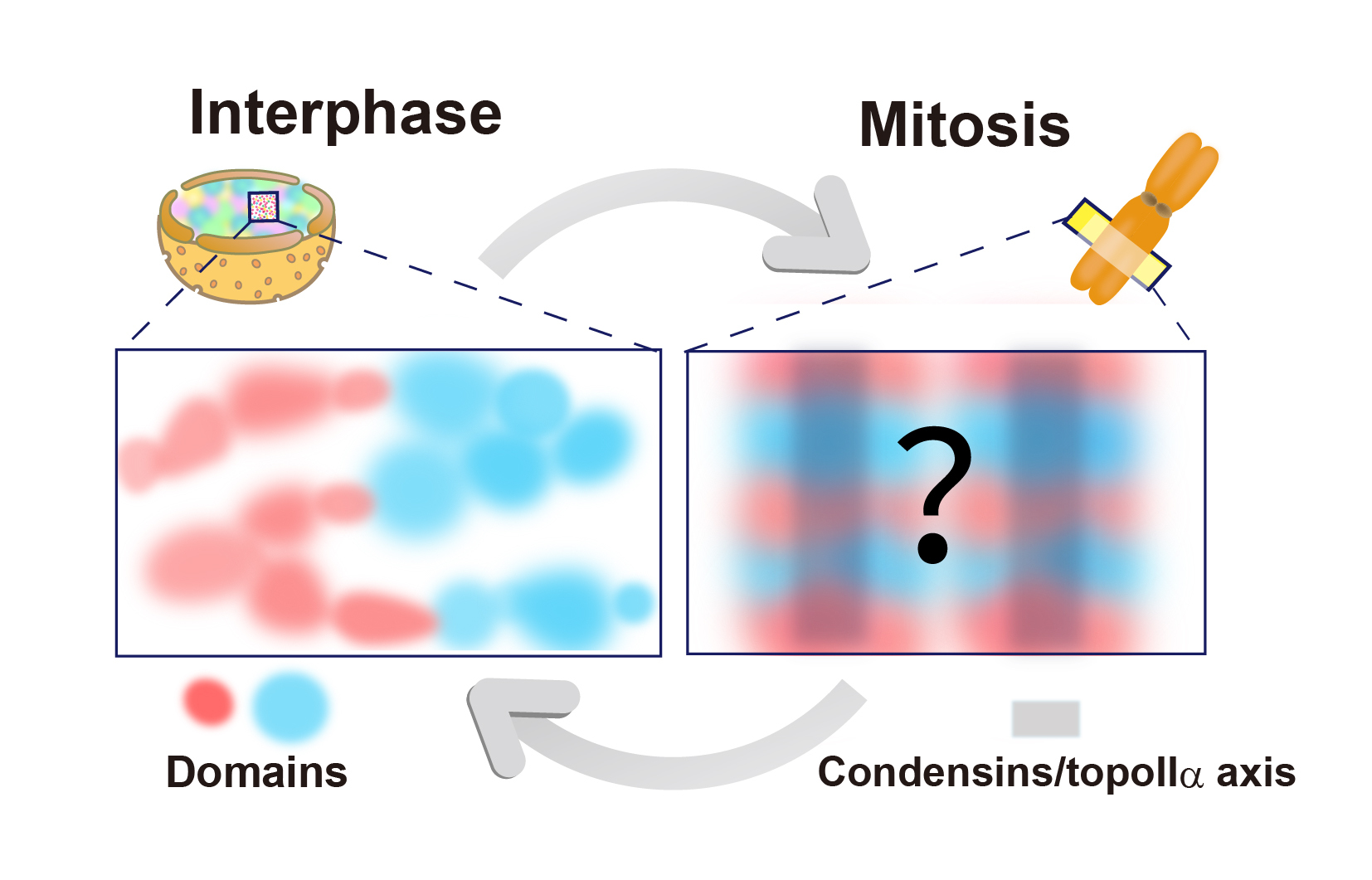
In interphase nuclei, chromatin is broadly divided into active euchromatic regions (red) and transcriptionally inactive heterochromatic regions (blue), but both exist as condensed chromatin domains (left). During mitosis, these pre-existing chromatin domains may be gathered, crosslinked, and reorganized by condensins, topoisomerase IIα, electrostatic interactions, free Mg2+, depletion attraction, and other factors to form mitotic chromosomes (right).