
Symbiosis and Cell Evolution Laboratory
Sexual life cycle establishes the unicellular red algae Cyanidiophyceae as a genetically tractable model for eukaryotic evolution.
Shunsuke Hirooka, Takayuki Fujiwara, Mark Seger, Soichi Inagaki, Shota Yamashita, Dai Tsujino, Ryo Onuma, Yu Kanesaki, Satoru Watanabe, Yuu Hirose, Ryudo Ohbayashi, Mari Takusagawa, Baifeng Zhou, Reiko Tomita, Fumi Yagisawa, Peter Lammers, Atsuko H Iwane, Shin-ya Miyagishima.
The Plant Cell 38(7): koag148, 2026 DOI:10.1093/plcell/koag148
This paper was featured in the “In Brief” section of the July 2026 issue of The Plant Cell and was highlighted in a commentary article by Dr. Matteo Pivato titled “The hidden life of unicellular red algae: discovery of a common sexual cycle unlocks a model lineage for eukaryotic evolution.”
We elucidated sexual life cycles, alternating between cell-walled diploid and cell wall-less haploid phases, in all cultivable genera of the unicellular red algal class Cyanidiophyceae, an early-diverging lineage within Archaeplastida comprising red algae, glaucophytes, green algae, and land plants. In addition, we generated high-quality genomic resources with phase-specific transcriptomes and developed genetic manipulation systems using the cell-wall-less haploids of these genera. We further uncovered phase-specific distribution of histone H3 lysine 27 trimethylation linked to haploid- and diploid-specific gene expression, including transcription factors involved in differentiation associated with sexual reproduction in plants. Together, these advances establish Cyanidiophyceae as a powerful model lineage for studying the evolution of sex, life cycles, epigenetic regulation, and environmental adaptation in Archaeplastida, with broader implications for eukaryotic evolution.
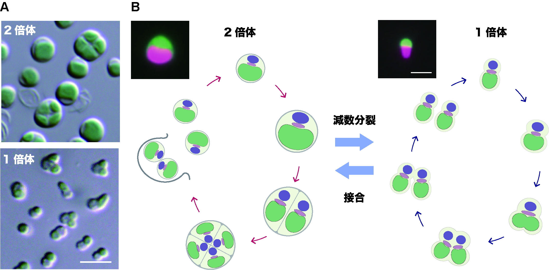
Sexual life cycle identified in Cyanidiophyceae. (A) Light micrographs of diploid (upper) and haploid (lower) Cyanidioschyzon merolae cells. Scale bar: 5 μm. (B) Schematic representation of the sexual life cycle in cyanidiophycean algae. Fluorescence images at the top show cells expressing the green fluorescent protein Venus in the cytoplasm. Scale bar: 2 μm.