


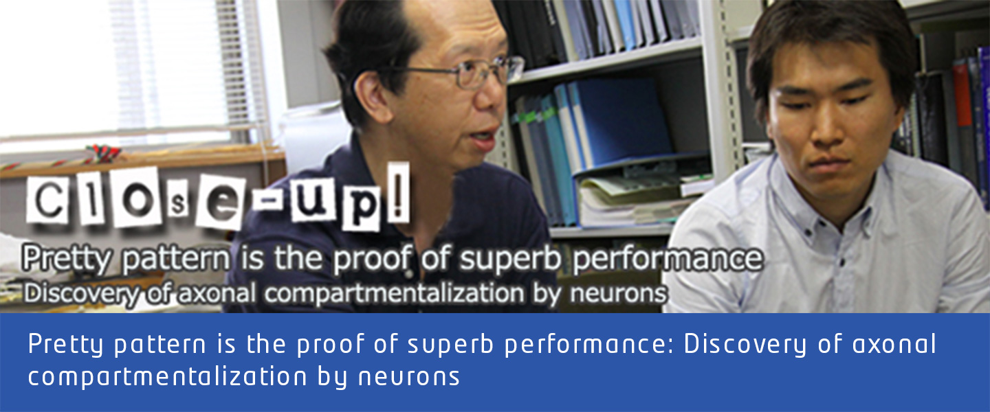
| Yasushi HIROMI, D. Sc., Professor / Takeo KATSUKI, Postdoc Hiromi Group, Division of Developmental Genetics, Department of Developmental Genetics, National Institute of Genetics/ SOKENDAI. |
The human brain is believed to have 100 to 200 billion neurons. Neural circuits composed of neurons regulate behavior, cognition, emotion, and the maintenance of life itself. Neurons develop long protrusions called axons, which act as “wires” in the formation of neural circuits and serve as “cables” for information transmission within the circuits. Neural circuit formation and function depend on various cellular properties of neurons. Studies have revealed that proteins found on the membrane surface are different at the root and the tip of the axon, but the mechanism behind this intra-axonal distribution has been a mystery. Prof. Yasushi HIROMI of the Division of Developmental Genetics, Department of Developmental Genetics and his research team including Researcher Dr. Takeo Katsuki have recently discovered that such localized protein distribution is due to the compartmentalization of the axon into two distinct regions. They have named this phenomenon “intra-axonal patterning.
Katsuki: “It is pretty.”
Hiromi: “That is to say, we demonstrated exactly what many researchers had long wished to see. People knew that membrane molecules could be localized within the axonal membrane, but had only seen such a phenomenon in bundle of axons, and had never observed the distribution within a single axon. Some had predicted that intra-axonal distribution would be destroyed when individual axons are separated from each other. In our research, we allowed neurons dissociated from the embryo to grow axons in the culture dish, and then visualized the distribution of membrane proteins. We obtained images indicating that even when axons were separated from its normal environment, specific proteins are still localized to segments of axons: one protein labeled with green were localized from the root to the middle, and another labeled with red from the middle to the tip of the axon. We were quite impressed with this phenomenon.”
Dr. Hiromi and his team used the neurons of Drosophila embryos in their research. The proteins labeled red and green are called “axon guidance receptors” and are involved in steering the direction of axonal growth during the formation of neural circuits. The distribution of axon guidance receptors was previously believed to result from the temporally-restricted pattern of expression during axon elongation. Dr. Katsuki and his colleagues designed experiments to challenge this hypothesis.
Hiromi: “There were two possibilities: Let’s say there are two axon guidance receptors, A and B. One possibility is “temporal control”, as previously been thought: in the early phase of axonal development only A is produced, and then at a certain point in time switches to the production of B. The other possibility is a “spatial control” that is not influenced by the timing of production of A or B. In other words, A and B are placed at right places where they are supposed to be.”
Katsuki: “To verify the first possibility, we artificially controlled the timing of production of A and B. But no matter what we did, they appeared at the right places. This demonstrated that timing is not important. On the other hand, when we tampered with the trafficking of intracellular material, their location was disturbed. This suggests the important role played by “intracellular logistics” (material transportation route) in intra-axonal patterning. Perhaps molecules such as A and B have something like “address labels” and a system exist that reads such addresses and delivers them correctly to the final destination.”
Hiromi: “That being said, the metaphor of logistics as in the human society can’t fully apply to cells. For example, the axon membrane is made of lipid, which is highly fluid. Since membrane proteins “float” on the lipid layer and can move around, membrane proteins could disperse and eventually end up completely mixed, even if different proteins are initially delivered to their correct locations. There must be a mechanism that keeps the proteins confined to specific axonal segments. We used a technology that enables us to observe the mobility of proteins in live neurons, and identified a “barrier” in the membrane that can prevent molecular dispersion, near the center of the axon. We believe that this barrier divides the axon into compartments to keep proteins at the right places. Trafficking and barrier work in tandem, so to speak.”
Since intra-axonal patterning is observed in more than 90% of neurons from the Drosophila embryo, it is believed to be a fundamental property of neurons. Moreover, intra-axonal protein distribution similar to that of Drosophila neurons have also been reported from mouse tissues, suggesting that it is highly likely that the patterning mechanism that the NIG team has discovered also exists in mouse and human.
Dr. Hiromi’s laboratory has obtained data that suggest axonal compartmentalization constitutes the foundation of neural circuit formation and higher functions. For example, the laboratory has shown that receptors on the axon surface capture secreted factors and localize them within the tissue, thus providing them as “guideposts” for axonal growth by neurons.
“Neurons have turned out to be far more sophisticated than we used to imagine,” says Dr. Hiromi. In future, it may be possible to find a link between abnormal intra-axonal patterning and serious neurological diseases.
“To do so, we must first elucidate the molecular mechanism of axonal compartmentalization,” says Dr. Katsuki. Another tasks for the future is to find out if a similar patterning mechanism can be found in axons in live organisms, not just cultured cells.
Katsuki: “That’s where we can make put the “Awesome power of Drosophila genetics” to the maximum use. In the future, when we carry out similar analyses in vivo, I’m sure we can obtain far prettier and more impactful results.”
Further elucidation of the mechanism of intra-axonal patterning is eagerly awaited.
“I worked in Dr. Hiromi’s laboratory for about one month as an intern student when I was an undergraduate student. During this period, I was seriously ‘imprinted’ by him! I then joined the lab as a graduate student and have since been doing my research in this laboratory. I learned to find joy in the pursuit of truth, even if that may take a long time — I believe this is the essence of science.”
(Interviewed by Naoko Nishimura on Sep. 7, 2009)
Takeo Katsuki, Deepak Ailani, Masaki Hiramoto and Yasushi Hiromi.Neuron, Vol. 64, Issue 2, 188-199, 2009. DOI: 10.1016/j.neuron.2009.08.019
Back


